EN
EN- DE
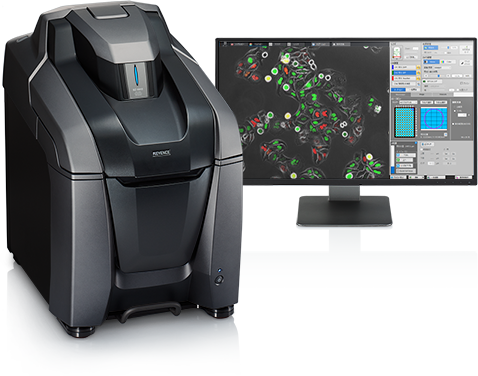
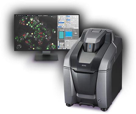
Capture publication-quality images quickly and easilyClear Fluorescence, Bright Field and Phase Contrast ImagesCompact Fluorescence Microscope BZ-X800


Optical Sectioning Using Structured Illumination
Optical sectioning provides clear images without fluorescence blurring.


Tubulin and H2AX Courtesy of Momoko Ishikawa, Department of Pediatric Dentistry, Tohoku University Graduate School of Dentistry
Capturing Wide Fields in High-Resolution and High-Speed
Up to 50.000 x 50.000 pixels can be quickly joined together.
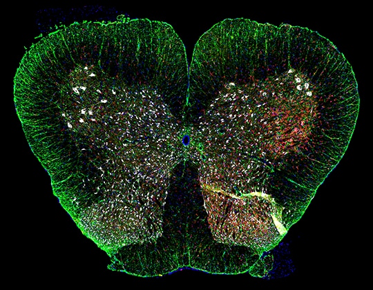
Rat spinal cord Courtesy of Professor Tasuku Nishihara, Department of Anesthesia and Perioperative Medicine, Ehime University Graduate School of Medicine
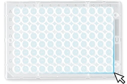
Image Cytometry for Visible Quantification
- 01Quickly scan an entire well-plate
- 02High-content analysis with uniform conditions
- 03Automatically batch capture desired images
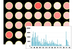
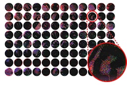
Capture and analyse large data sets in a short time.
Clear images provide precise quantification.
Accurate Analysis of 3D Localization
Quantification conditions apply quickly to an entire Z-stack.
3D Analysis
Motion Analysis
Movements of objects caused by morphological changes can be tracked.
Data, such as signal intensities or surface areas, can then be output.
Further details and application examples can be found in the following brochures.
- Kontaktieren Sie uns unter: